The Pathobyte Series: Treponema pallidum- The Microscopic Stealth Drill
First identified in 1905, Treponema pallidum is a versatile Gram-indeterminate bacterium capable of causing the devastating human illness syphilis. Driven by an advanced arsenal of internal endoflagella and a stealthy outer membrane, it effortlessly evades host immunity, spreading rapidly through direct, close human contact. Infections range from superficial, painless sores to life-threatening systemic conditions, including severe neurological and deep organ damage. Accurate identification leverages classic antibody blood tests like the Wassermann assay and modern tools like long-term rabbit-cell cultivation. While treatment typically requires targeted antimicrobial chemotherapy, robust prevention relies heavily on cutting-edge genetic transformation mapping and proactive vaccine design.
How Did a Pale, Invisible "Turning Thread" Terrify Ancient Europe?
Long ago, before doctors had microscopes or modern medicines, this shape-shifting germ caused a terrifying, widespread disease called syphilis. It first exploded across Europe in 1495, spreading panic because it caused painful sores and deep bone aches. Because nobody knew where it came from, countries blamed each other! The French called it the "Neapolitan disease," while the English called it the "French disease." People even blamed the alignment of planets or angry gods because they did not have any scientific explanations.
For over four hundred years, the true cause of this illness was a complete mystery to doctors. Finally, in 1905, two scientists named Fritz Schaudinn and Erich Hoffmann spotted the culprit under a microscope. It was so pale and thin that it was nearly invisible. They named it Treponema pallidum, which comes from the Greek and Latin words that mean turning thread and pale. This discovery completely changed how scientists studied diseases because they finally had a real target to look at and study under their microscopes.
Today, we know this tiny organism is a specialized type of bacterium that can live inside humans for decades without being seen. When it first enters a person, it creates a small, painless sore at the entryway. If it is not treated by a doctor, the germ slowly travels to other organs and can cause massive damage over time. It is a slow-moving invader, but its ability to hide makes it one of the most successful and dangerous microscopic hitchhikers in human history.
Why Does This Fragile Parasite Need a Human Body to Stay Alive?
This microbe is an obligate parasite, which means it cannot survive on its own in the outside world. It does not live on doorknobs, soil, or water like other common germs. It needs a warm, cozy human host to stay alive. Because it has lost almost all its internal machinery through evolutionary genome reduction, it relies completely on our bodies to get its food. Without a human home, this fragile germ dries out and dies within just a few minutes of leaving a body.
Because it cannot live on surfaces, it spreads through direct, close human-to-human contact. Once it enters a new host, it uses its unique body shape to navigate through body fluids easily. It loves to slide into the bloodstream and travel to different areas. Instead of staying in one place, it spreads out quickly to find the best spots to set up camp. This fast movement helps it escape the initial defensive reactions that the human body throws against new invaders.
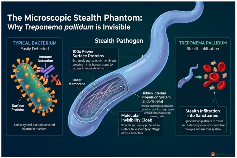
It loves to hide out in safe zones like the eyes or the nervous system, where the human immune system struggles to reach. Scientists call these places protected sanctuaries because the body limits its own defensive attacks there to prevent self-damage. By slipping into these quiet areas, the germ can rest and multiply safely for years. It avoids the heavy artillery of our body defenses, turning a quick infection into a lifelong game of hide-and-seek that requires powerful medicine to stop.
How Does This Living Corkscrew Drill Through Our Internal Defenses?
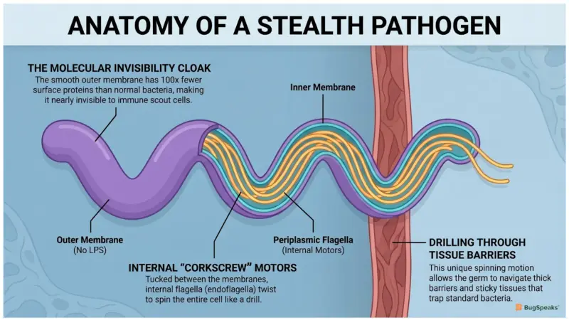
Treponema pallidum is a true master of stealth because of its unique skin structure. It is wrapped in a smooth outer membrane that has almost no identifying markers on it. Scientists call it a stealth pathogen because it wears a molecular invisibility cloak, making it hard for our defense forces to spot it. While normal bacteria have lots of proteins sticking out of their skin like flags, this germ keeps its surface clean so it looks just like normal human tissue.
To move around, it uses special internal motors called endoflagella that are tucked away safely inside its walls. Instead of flapping long tails outside its body, these hidden ropes twist inside its layers, causing the whole bacterium to spin like a drill. It uses this powerful corkscrew motion to tear through sticky tissues and slide into blood vessels easily. This allows it to swim through thick barriers that would easily trap and immobilize standard bacteria with external tails.
Once inside the bloodstream, the germ interacts with human platelets, which are the tiny cells that help clot your blood. It grabs onto these cells to hitchhike across the body and stick to blood vessel walls. This interaction changes how the bacterium moves and helps it exit the bloodstream into target tissues. By hijacking these cells, it travels anywhere it wants, slipping through tiny gaps in blood vessels to colonize deep organs before the body can even launch a proper defensive counter-attack.
[Germ enters through skin] ──► [Uses endoflagella to drill] ──► [Hijacks blood platelets] ──► [Hides from immune system]
How Did Fighting This Microbe Launch the Era of Magic Bullets and Fever Cures?
While the germ itself is dangerous and provides no direct health benefits, our fight against it has completely changed modern medicine. In 1910, scientists created a special compound called Salvarsan to fight this germ. This was the birth of modern antimicrobial chemotherapy, which means using specific chemicals to hunt down a single bacterium. Before this discovery, people used toxic metals that made patients sicker, so learning how to build targeted chemical magic bullets saved millions of lives from various infections around the world.
Even more surprising, a doctor named Julius Wagner-Jauregg noticed this germ hates heat because it completely lacks cellular alarms to protect itself from high temperatures. He intentionally gave infected patients malaria to trigger high fevers that cooked the fragile bacteria inside the brain. Once the bacteria died, he cured the malaria with medicine. This bizarre treatment, called pyrotherapy, brought a fatal brain condition under clinical control and won the Nobel Prize in 1927, proving that understanding a germ's weakness can inspire radical cures.
Developing early diagnostics for this germ also catalyzed major progress in clinical immunology, which is the study of how our bodies fight off invaders. The famous Wassermann test, invented in 1906, was the first widely used blood test to look for signs of an infectious disease. It taught scientists how to use blood proteins to check if someone was sick before symptoms even appeared. Our long history of fighting this single corkscrew germ essentially built the foundations of modern drug discovery, immune testing, and hospital laboratories.

How Are Modern Genetic Blueprints Helping Us Create the Ultimate Shield?
Even today, this microscopic drill is a major global health focus for scientists everywhere. For over a hundred years, researchers could not grow it in a regular laboratory dish because it would instantly die without a host. It was not until 2018 that scientists finally figured out a long-term cultivation system using rabbit cells and a low-oxygen atmosphere. Being able to keep this germ alive in a lab dish finally opened the door for deep genetic research that was completely impossible for past generations.
This lab breakthrough allowed scientists to perform genetic transformation on the bacterium for the very first time in history. By cutting out specific genes, researchers are finally figuring out which proteins the germ uses to build its invisibility cloak. This helps us find new drug targets and understand how the germ behaves. It is a massive step forward because understanding the exact genetic blueprint of an enemy is the only way to find its permanent weakness and stop it from spreading.
Now, scientists are using these genetic tools to design a modern vaccine that can train our immune systems to spot the stealth invader instantly. Because the germ is slowly becoming resistant to older medicines in some parts of the world, having a preventative shield is more important than ever. The lessons we are learning from this ancient shape-shifter are helping us prepare for future medical challenges. The tiny turning thread continues to push human science forward into a brand new era.
Taxonomic Classification
Microbe Profile
Shape: Helical, long, slender, and spiral-shaped (looks like a corkscrew).
Gram Stain Nature: Structurally Gram-negative, but clinically Gram-indeterminate because it is too thin to absorb standard dyes.
Spore-Forming: Non-spore-forming.
Biofilm Formation: Non-biofilm-forming on its own.
Oxygen Requirement: Microaerophilic (requires a tiny amount of oxygen, between 1.5% and 5%).
Optimal Temperature: Cool! Prefers 33°C to 35°C. It dies quickly if temperatures reach 41.5°C.
Optimal pH: 7.2 to 7.4 (neutral, matching our body fluids).
Nutrient Usage: Chemoorganoheterotroph. It cannot make its own food and steals sugars, amino acids, and fats directly from its human host.
Fun Facts
The Stealth Phantom: Its outer membrane has 100 times fewer surface proteins than standard bacteria, making it practically invisible to our body’s scout cells!
Platelet Hijacker: It literally grabs onto human blood clotting cells (platelets) to hitchhike through the bloodstream.
Biochemically Helpless: It completely lacks heat-shock proteins. If it gets too hot, its internal enzymes simply break apart, leaving it defenseless against a high fever.
-Varsha V
Reference
Vrioni, G., & Peppas, T. A. (2024). What’s in a Name? Hellenic Origins of Microbiological Nomenclature. Acta Microbiologica Hellenica, 69(2), 93-100. https://doi.org/10.3390/amh69020010
Gantenbein U. L. (2025). The poet Ulrich von Hutten (1488-1523) and the French disease: the records and human remains of a probable yaws patient. Medical history, 69(1), 1–21. https://doi.org/10.1017/mdh.2024.38
Edmondson, D. G., Hu, B., & Norris, S. J. (2018). Long-Term In Vitro Culture of the Syphilis Spirochete Treponema pallidum subsp. pallidum. mBio, 9(3), e01153-18. https://doi.org/10.1128/mBio.01153-18
Houston, S., Schovanek, E., Conway, K. M. E., Mustafa, S., Gomez, A., Ramaswamy, R., Haimour, A., Boulanger, M. J., Reynolds, L. A., & Cameron, C. E. (2022). Identification and Functional Characterization of Peptides With Antimicrobial Activity From the Syphilis Spirochete, Treponema pallidum. Frontiers in microbiology, 13, 888525. https://doi.org/10.3389/fmicb.2022.888525
Norris S. J. (2025). Establishment of the Nichols strain as the type strain of Treponema pallidum. International journal of systematic and evolutionary microbiology, 75(2), 006697. https://doi.org/10.1099/ijsem.0.006697
Phan, A., Romeis, E., Tantalo, L., & Giacani, L. (2022). In Vitro Transformation and Selection of Treponema pallidum subsp. pallidum. Current protocols, 2(8), e507. https://doi.org/10.1002/cpz1.507
Zobaníková, M., Mikolka, P., Cejková, D., Pospíšilová, P., Chen, L., Strouhal, M., Qin, X., Weinstock, G. M., & Smajs, D. (2012). Complete genome sequence of Treponema pallidum strain DAL-1. Standards in genomic sciences, 7(1), 12–21. https://doi.org/10.4056/sigs.2615838
Ng, H. M., Slakeski, N., Butler, C. A., Veith, P. D., Chen, Y. Y., Liu, S. W., Hoffmann, B., Dashper, S. G., & Reynolds, E. C. (2019). The Role of Treponema denticola Motility in Synergistic Biofilm Formation With Porphyromonas gingivalis. Frontiers in cellular and infection microbiology, 9, 432. https://doi.org/10.3389/fcimb.2019.00432
Buyuktimkin, B., Zafar, H., & Saier, M. H., Jr (2019). Comparative genomics of the transportome of Ten Treponema species. Microbial pathogenesis, 132, 87–99. https://doi.org/10.1016/j.micpath.2019.04.034
Knauf, S., Hisgen, L., Ågren, E. O., Barlow, A. M., Faehndrich, M., Voigt, U., Fischer, L., Grillová, L., Hallmaier-Wacker, L. K., Kik, M. J. L., Klink, J. C., Křenová, J., Lavazza, A., Lüert, S., Nováková, M., Čejková, D., Pacioni, C., Trogu, T., Šmajs, D., & Roos, C. (2024). High prevalence and genetic diversity of Treponema paraluisleporidarum isolates in European lagomorphs. Microbiology spectrum, 12(1), e0177423. https://doi.org/10.1128/spectrum.01774-23
Liu, J., Howell, J. K., Bradley, S. D., Zheng, Y., Zhou, Z. H., & Norris, S. J. (2010). Cellular architecture of Treponema pallidum: novel flagellum, periplasmic cone, and cell envelope as revealed by cryo electron tomography. Journal of molecular biology, 403(4), 546–561. https://doi.org/10.1016/j.jmb.2010.09.020