The Pathobyte Series: Diphtheria – Silent Throat Killer and Its Deadly Toxin
First isolated in 1884, Corynebacterium diphtheriae is a versatile Gram-positive bacterium capable of causing a wide array of human illnesses. Driven by an advanced arsenal of virulence factors and toxins like the diphtheria toxin, it effortlessly evades host immunity, spreading rapidly through respiratory droplets and close contact. Infections range from localized pharyngeal lesions featuring a leathery pseudomembrane to life-threatening systemic conditions, including myocarditis and polyneuropathy. Accurate diagnosis leverages classic laboratory cultures on selective tellurite media and modern tools like PCR testing. While treatment typically requires targeted antibiotic therapy using penicillin alongside urgent antitoxin administration for toxigenic strains, robust prevention relies heavily on routine toxoid immunization protocols.
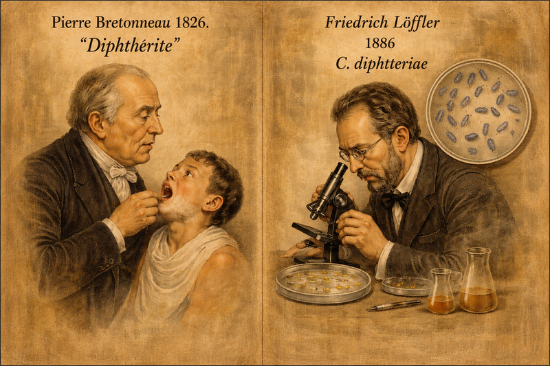
History
The clinical entity of diphtheria was first clearly described by the French physician Pierre Bretonneau in 1826, who coined the term 'diphthérite' from the Greek word for leather, referring to the characteristic tough throat membrane. Friedrich Löffler isolated the causative bacillus in 1884, now known as C. diphtheriae.
Pathogenesis
C. diphtheriae is a gram-positive, non-spore-forming, non-motile bacillus that colonises the upper respiratory tract. Its pathogenicity is driven almost entirely by diphtheria toxin (DT), encoded by the tox gene located on the lysogenic corynephage β. Only toxigenic strains carry this phage. The toxin is a two-fragment (A-B) exotoxin: the B fragment binds to the heparin-binding epidermal growth factor precursor on host cell surfaces, enabling receptor-mediated endocytosis, while the A fragment catalyses ADP-ribosylation of elongation factor-2 (EF-2), irreversibly halting eukaryotic protein synthesis. This mechanism underlies local pseudomembrane formation in the pharynx and systemic complications including myocarditis, polyneuropathy, and renal tubular necrosis. Locally, the organism elicits a fibrinous, inflammatory exudate forming the classic greyish-white pseudomembrane, tightly adherent and bleeding upon attempted removal.
Transmission
Diphtheria is transmitted primarily via respiratory droplets from infected individuals or asymptomatic carriers. Close, prolonged contact such as household exposure significantly elevates infection risk. Fomite transmission has also been documented, though less common. Cutaneous diphtheria, characterised by infected skin ulcers, can transmit through direct contact with wound secretions and is endemic in tropical and resource-limited settings. The incubation period typically ranges from 2 to 5 days (range: 1–10 days). Importantly, individuals may carry and transmit C. diphtheriae without exhibiting clinical disease, sustaining community transmission.
Signs and Symptoms
Clinical diphtheria typically manifests as pharyngeal/tonsillar diphtheria, the most common form, characterised by: Low-grade fever, sore throat, and malaise in the prodromal phase (1–2 days), Formation of a tough, greyish-white pseudomembrane over the tonsils/pharynx that bleeds on removal, 'Bull neck' appearance due to cervical lymphadenopathy and soft-tissue oedema, Hoarseness, stridor, and potential airway obstruction in laryngeal involvement (croup), Myocarditis (in ~10–25% of cases), presenting as arrhythmias or heart block, typically 1–2 weeks after onset, Neuropathy (palatal palsy, oculomotor palsy, peripheral motor neuropathy) weeks after illness onset. Cutaneous diphtheria presents as chronic, non-healing ulcers with a membranous base.

Diagnosis
Diagnosis is primarily clinical but must be confirmed microbiologically. Throat and nasopharyngeal swabs should be obtained before antibiotic initiation and cultured on selective media, Löffler's serum slope, and tellurite medium (e.g., Hoyle's medium), where C. diphtheriae produces characteristic grey-black colonies. Gram staining reveals gram-positive 'Chinese letter' or palisade arrangements. Toxigenicity must be confirmed using the Elek immunodiffusion test (gold standard) or PCR for the tox gene. The WHO recommends immediate treatment upon clinical suspicion without awaiting laboratory confirmation, given the rapid progression and high mortality.
Treatment
Treatment involves two parallel strategies: toxin neutralisation and bacterial eradication. Diphtheria Antitoxin (DAT), derived from equine serum, neutralises circulating (unbound) toxins and is the cornerstone of treatment; it must be administered promptly, as it cannot reverse toxins already bound to tissues. Dosing ranges from 20,000 to 100,000 IU intramuscularly or intravenously depending on disease severity, after testing for hypersensitivity. Antibiotics erythromycin (40–50 mg/kg/day orally for 14 days) or penicillin G eliminate the organism, terminate toxin production, and prevent carriage; penicillin is the preferred agent per WHO guidelines. Patients should be isolated, and supportive care, airway management, cardiac monitoring, and nutritional support are critical in severe cases. Contacts require prophylaxis with antibiotics and booster vaccination regardless of prior immunisation status.
Prevention
Vaccination is the most effective preventive measure. Diphtheria toxoid, formulated with tetanus toxoid and pertussis vaccine (DTP/DTaP/Tdap), is recommended by the WHO as part of the Expanded Programme on Immunisation. The primary series (3 doses) should begin at 6 weeks of age with booster doses at 18 months, 4–6 years, and in adolescence. Maintenance of >95% population coverage is needed to sustain herd immunity. In resource-limited settings or outbreak situations, mass immunisation campaigns are deployed. Surveillance systems for early case detection, strict isolation of confirmed cases, and contact tracing with chemoprophylaxis are essential public health tools. The WHO and CDC continue to report resurgences in regions with declining vaccination coverage, a stark reminder that diphtheria, though vaccine-preventable, remains an active threat wherever immunisation systems falter.
Microbial Profile
Gram nature: Gram-positive
Spore Formation: Non-spore forming
Motility: Non-motile
Shape: Rod-shaped
Oxygen Requirement: Aerobic
Optimal Temperature: 37℃
Optimal pH: 7.2–8.0
Taxonomic Classification
Domain: Bacteria
Kingdom: Bacillati
Phylum: Actinomycetota
Class: Actinomycetes
Order: Mycobacteriales
Family: Corynebacteriaceae
Genus: Corynebacterium
Species: Corynebacterium diphtheriae
-Varsha V
Reference
Holmes R. K. (2000). Biology and molecular epidemiology of diphtheria toxin and the tox gene. The Journal of infectious diseases, 181 Suppl 1, S156–S167. https://doi.org/10.1086/315554
Burkovski, A. (2015). Pathogenesis of Corynebacterium diphtheriae and Corynebacterium ulcerans . In Human Emerging and Re-emerging Infections, S.K. Singh (Ed.). https://doi.org/10.1002/9781118644843.ch38
Jamal, S. B., Tiwari, S., Silva, A., & Azevedo, V. (2017). Pathogenesis of Corynebacterium diphtheriae and available vaccines; an overview. Glob. J. Infect. Dis. Clin. Res, 3, 20-24.
Valenzuela-Cardenas, M. B., & Goodfellow, S. M. (2025). Microbe Snapshot: Corynebacterium diphtheriae. Clinical Microbiology Newsletter.
Ningsih, I., & Safrullah, M. I. (2025). Pathogenesis, Diagnosis, and Examination of Corynebacterium diphtheriae. Indonesian Journal of Medical Chemistry and Bioinformatics, 3(2), 1.
Murphy JR. Corynebacterium Diphtheriae. In: Baron S, editor. Medical Microbiology. 4th edition. Galveston (TX): University of Texas Medical Branch at Galveston; 1996. Chapter 32. Available from: https://www.ncbi.nlm.nih.gov/books/NBK7971/
Hoskisson, P. A. (2018). Microbe Profile: Corynebacterium diphtheriae–an old foe always ready to seize opportunity. Microbiology, 164(6), 865-867.