The PathoBytes Series: Trichomonas vaginalis: The Parasite Behind Non-Viral STI
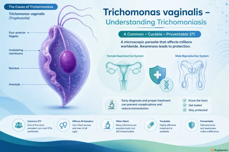
This blog explores Trichomonas vaginalis, a microscopic protozoan parasite responsible for trichomoniasis, the world's most common non-viral sexually transmitted infection (STI). Although millions of new infections occur every year, the disease often goes undiagnosed because many infected individuals have few or no symptoms. Beyond causing vaginal discharge, irritation, and urethritis, T. vaginalis is associated with adverse pregnancy outcomes, infertility, and an increased risk of acquiring and transmitting HIV. This article examines the parasite's biology, transmission, pathogenesis, clinical manifestations, diagnosis, treatment, prevention strategies, and the ongoing public health challenges in controlling this widespread yet frequently overlooked infection.
What is Trichomonas vaginalis?
Trichomonas vaginalis is an obligate human parasite that exists exclusively as a mobile trophozoite; unlike many other protozoans, it lacks a resistant cyst stage in its life cycle. The parasite exhibits a characteristic teardrop or pear-shaped morphology and utilizes four anterior flagella along with an undulating membrane to move with a distinct "jerky" or tumbling motility.
As an extracellular parasite, Trichomonas vaginalis adheres directly to the epithelial lining of the urogenital tract. In women, the primary site of infection is the squamous epithelium of the vagina and ectocervix. In men, the organism colonizes the mucosal surfaces of the urethra, anterior prostate, and seminal vesicles. The resulting condition, trichomoniasis, is characterized by mucosal inflammation, cellular destruction, and altered vaginal microecology.
Public Health Significance and Global Prevalence
The public health impact of Trichomonas vaginalis extends far beyond localized genital discomfort:
Epidemiological Weight: It accounts for nearly half of all curable non-viral STI cases worldwide.
HIV Co-factor: The intense inflammatory response and mucosal breakdown caused by T. vaginalis significantly increase the likelihood of acquiring or transmitting HIV.
Reproductive Morbidity: In pregnant women, untreated infection is associated with premature rupture of membranes (PROM), preterm labor, and low birth weight infants.
Asymptomatic Carriage: Up to 70% of infected individuals are asymptomatic, creating a large, undetected reservoir that drives ongoing community transmission.
History and Naming
Discovery and Scientific Milestones
In 1836, French physician and microscopist Alfred François Donné observed motile, flagellated microorganisms in purulent vaginal secretions from women suffering from vaginitis. He initially designated the organism Infusorie vaginal.
Recognizing the distinct morphology of the parasite, French zoologist Félix Dujardin subsequently assigned it to a new genus, coining the name Trichomonas vaginalis in 1837.
Origin of the Name
The scientific name reflects the morphological and anatomical characteristics of the organism:
"Tricho-": Derived from the Greek word thrix (genitive trichos), meaning hair, referring to the whip-like flagella.
"-monas": Derived from the Greek monas, meaning a single unit or simple unicellular organism.
"vaginalis": Refers to the primary site of discovery and colonization in human hosts—the vagina.
Microbe Profile
The following profile details the physical, metabolic, and physiological characteristics of Trichomonas vaginalis:
Taxonomic Classification
The taxonomic hierarchy of Trichomonas vaginalis according to modern eukaryotic phylogenetic classifications is presented below:
Pathogenesis
Virulence Factors and Adhesion Mechanisms
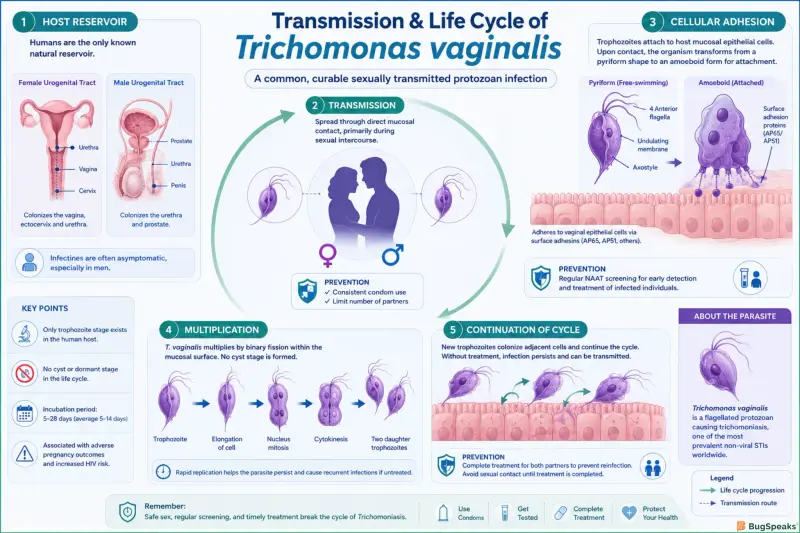
Upon entering the urogenital tract, Trichomonas vaginalis undergoes a rapid transformation from a free-swimming pyriform trophozoite into a flattened, amoeboid form. This morphological change increases the parasite's surface area, allowing it to contact host epithelial cells directly.
Key molecular factors driving adhesion and colonization include:
Adhesion Proteins (APs): Surface proteins including AP65, AP51, AP33, and AP23 mediate specific receptor-binding interactions with fibronectin and laminin on human vaginal epithelial cells (VECs).
Lipoglycan (TvLG): The major cell-surface glycoconjugate of T. vaginalis, TvLG binds to host galectin-1 and galectin-3 receptors, initiating inflammatory signaling and anchoring the parasite to host tissue.
Cysteine Peptidases (CPs): The parasite secretes over 20 distinct cysteine proteases (such as TvCP39) that degrade host extracellular matrix proteins, immunoglobulins (IgA and IgG), and complement components.
Cytotoxic Effects and Host Inflammatory Response
Once attached, T. vaginalis exerts direct cytotoxic actions on host cells through nutrient depletion, mechanical disruption, and the release of pore-forming proteins (perforin-like factors). The parasite actively phagocytoses host erythrocytes, vaginal epithelial cells, and commensal lactobacilli.
The host immune system responds by recruiting neutrophils, macrophages, and lymphocytes to the mucosal surface. Neutrophil infiltration generates reactive oxygen species (ROS) and pro-inflammatory cytokines. This intense local inflammatory cascade causes epithelial sloughing, micro-ulcerations, and small petechial hemorrhages.
Immune Evasion and Parasite Survival
Trichomonas vaginalis employs several mechanisms to persist within the host despite an active immune response:
Antibody Cleavage: Surface-bound and secreted cysteine peptidases degrade host secretory IgA and IgG antibodies before they can opsonize the parasite.
Phenotypic Variation: The parasite dynamically alters its surface antigen expression in response to environmental stressors and host immune pressure.
Phenotypic Masking:T. vaginalis absorbs host plasma proteins (such as human serum albumin and immunoglobulins) onto its outer membrane, concealing its foreign antigens from host immune surveillance.
Transmission
Modes of Transmission
Sexual Transmission: The predominant mode of transmission is unprotected sexual intercourse (vaginal-penile, vaginal-vaginal, or oral-genital contact). Transmission rates are high; a single sexual exposure with an infected partner carries a substantial risk of acquiring the parasite.
Vertical Transmission: Transmission from an infected mother to a newborn during passage through an infected birth canal occurs in up to 5% of deliveries, potentially leading to neonatal respiratory tract or vaginal infections.
Non-Sexual Transmission:Non-sexual transmission via shared damp items (such as unwashed bath towels, wet sponges, or contaminated gynecological instruments) is extremely rare because the parasite lacks a protective cyst stage and degrades rapidly outside the human body.
Signs and Symptoms
The incubation period for Trichomonas vaginalis typically ranges from 5 to 28 days following exposure, although many individuals harbor the organism for months or years without developing clinical symptoms.
Clinical Presentation in Women
Approximately 50% to 70% of infected women experience symptoms, which may range from mild irritation to severe, acute vulvovaginitis:
Vaginal Discharge: Classic discharge is diffuse, thin, homogeneous, frothy, yellow-green in color, and possesses a strong, unpleasant odor.
Pruritus and Irritation: Severe itching, burning, and soreness of the vulva and vagina.
Dysuria: Pain or burning sensation during urination caused by urethral involvement.
Dyspareunia: Painful sexual intercourse stemming from vaginal inflammation.
Strawberry Cervix (Colpitis Macularis): Marked punctate hemorrhagic spots on the ectocervix, visible during speculum examination in approximately 2% to 5% of symptomatic women.
Clinical Presentation in Men
Up to 70% to 80% of infected men are asymptomatic carriers. When symptoms occur, they are often transient and mild:
Urethritis: Mild urethral discharge (purulent or clear) and urethral irritation.
Dysuria and Burning: Stinging or burning sensation during or immediately after ejaculation or urination.
Complications: Rare secondary complications include epididymitis, prostatitis, and reduced sperm motility leading to temporary subfertility.
Pregnancy Complications and HIV Link
Pregnancy Outcomes:T. vaginalis infection during pregnancy is associated with adverse maternal and fetal outcomes, including premature rupture of membranes (PROM), preterm birth (before 37 weeks), and low birth weight.
HIV Susceptibility: Micro-ulcerations and localized neutrophil infiltration recruit CD4+ T-lymphocytes and macrophages to the genital mucosa. This tissue damage increases susceptibility to HIV acquisition by 1.5- to 2-fold and elevates viral shedding in HIV-positive individuals.
Diagnosis
Clinical signs alone are insufficient to diagnose trichomoniasis because symptoms overlap significantly with bacterial vaginosis, vulvovaginal candidiasis, and chlamydial infection. Objective laboratory testing is essential.
Diagnostic Modalities
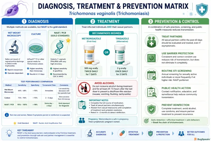
Wet Mount Microscopy:
Direct microscopic evaluation of fresh vaginal or urethral secretions suspended in 0.9% saline.
Mechanism: Visualization of motile, pear-shaped trophozoites displaying characteristic jerky movement.
Limitations: Low sensitivity (40%–60%); requires immediate evaluation (within 10–20 minutes of collection) before parasites lose motility.
Culture:
Historically considered the gold standard before molecular testing.
Mechanism: Inoculation into Specialized media (e.g., Diamond's medium, InPouch TV system) incubated at 37°C for 2–7 days.
Nucleic Acid Amplification Tests (NAATs):
Currently recommended as the gold standard diagnostic methodology by the CDC and WHO.
Mechanism: Amplification of parasite-specific RNA or DNA target sequences from vaginal swabs, endocervical swabs, or urine.
Performance: Exceptional sensitivity (95%–100%) and specificity
Rapid Antigen Tests & Point-of-Care (POC) Tests:
Immunochromatographic membrane assays (e.g., OSOM Trichomonas Rapid Test) that detect parasite surface antigens within 10–45 minutes.
Sensitivity: 80%–90%; useful in resource-limited clinical settings.
Comparison of Diagnostic Methods
Treatment
Systemic therapy is mandatory to eradicate Trichomonas vaginalis. Topical intravaginal creams or gels (such as metronidazole gel) are ineffective because they fail to reach therapeutic drug concentrations in infected urethral, skene gland, or prostatic tissue reservoirs.
First-Line Pharmacological Regimens
The 5-nitroimidazole class of drugs, specifically Metronidazole and Tinidazole, forms the backbone of trichomoniasis therapy.
Non-Pregnant Females (CDC Updated Guidelines):
Recommended Regimen:Metronidazole 500 mg orally twice daily for 7 days. (Superior cure rates compared to single-dose therapy).
Males:
Recommended Regimen:Metronidazole 2 g orally in a single dose, OR Metronidazole 500 mg twice daily for 7 days.
Alternative Regimen:
Tinidazole 2 g orally in a single dose. (Often better tolerated with fewer gastrointestinal side effects).
Partner Treatment and Management
Simultaneous treatment of all sexual partners is essential to prevent reinfection ("ping-pong" transmission). Patients must be instructed to abstain from sexual activity until both they and their partners have completed their antimicrobial course and are symptom-free (typically 7 days post-treatment).
Drug Resistance and Follow-up Testing
Nitroimidazole Resistance: Low-level metronidazole resistance occurs in approximately 4% to 10% of clinical isolates. Refractory cases are managed using higher doses of oral tinidazole (e.g., 2 g daily for 7 days) combined with high-dose metronidazole or specialized compound formulations.
Re-testing Recommendation: Due to high rates of post-treatment reinfection, re-testing via NAAT is recommended for all sexually active women within 3 months following initial therapy.
Prevention
Preventing trichomoniasis relies on combining barrier precautions, regular STI screening, prompt case identification, and partner notification.
Consistent Condom Use: Correct and consistent use of male latex or polyurethane condoms during every vaginal intercourse session significantly reduces the risk of T. vaginalis transmission.
Routine STI Screening: Annual screening is recommended for high-risk populations, including HIV-positive individuals, women entering correctional facilities, and patients attending STI clinics.
Simultaneous Partner Management: Expedited Partner Therapy (EPT) or direct partner referral ensures that partners receive concurrent treatment, preventing reinfection cycles.
Avoiding Unnecessary Douching: Discouraging vaginal douching preserves normal Lactobacillus microflora and maintains a protective acidic vaginal environment.
Vaccines
Current Status and Vaccine Challenges
There is currently no licensed human vaccine available for Trichomonas vaginalis. Developing an effective vaccine faces several biological hurdles:
Antigenic Variability:T. vaginalis strains display high phenotypic variation in surface proteins, allowing the parasite to escape antibody-mediated clearance.
Mucosal Immune Barriers: Eliciting long-lasting secretory IgA and cell-mediated immune responses within the lower genital tract mucosal tissue is difficult.
Short-Lived Natural Immunity: Natural infection does not confer long-term protective immunity; individuals can become reinfected immediately after successful antimicrobial cure.
Reference
World Health Organization. (2021). Global progress report on HIV, viral hepatitis and sexually transmitted infections. Geneva: World Health Organization.
Centers for Disease Control and Prevention. (2021). Sexually Transmitted Infections Treatment Guidelines: Trichomoniasis. MMWR Recommendations and Reports, 70(4), 1-187.
Poole, D. N., & McClelland, R. S. (2013). Global epidemiology of Trichomonas vaginalis. Sexually Transmitted Infections, 89(6), 418-422.
Kissinger, P. (2015). Epidemiology and treatment of Trichomonas vaginalis. Current Infectious Disease Reports, 17(6), 484.
Meites, E., Gaydos, C. A., Hobbs, M. M., et al. (2015). A review of evidence-based care for Trichomonas vaginalis infections. Clinical Infectious Diseases, 61(S8), S837-S843.
Hirt, R. P., & Sherrard, J. (2015). Trichomonas vaginalis origins, genomics and pathogenesis. Sexually Transmitted Infections, 91(4), 229-235.
Secor, W. E. (2012). Trichomonas vaginalis: the rogue protozoan. Journal of Parasitology Research, 2012, 730759.
Schwebke, J. R., & Burgess, D. N. (2004). Trichomoniasis. Clinical Microbiology Reviews, 17(4), 794-803.