The Pathobyte Series: Helicobacter pylori- The Stomach's Tiny Secret Agent
The micro-explorer blog uncovers Helicobacter pylori, a pale, spiral-shaped bacterium that survived undetected in the human stomach for thousands of years. Defying early medical beliefs that our digestive acids rendered the stomach sterile, this resilient microbe uses a special enzyme shield to neutralize burning acid. Equipped with whip-like tails, it drills into our protective stomach lining to build sticky biofilm fortresses. While this ancient organism can protect children from allergies, it also causes painful ulcers. Today, advanced genetic tools track its path through human history, while scientists transform its machinery into life-saving vaccines and targeted medical treatments.
How Did a Pale, Invisible "Turning Thread" Terrify Ancient Europe?
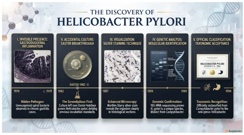
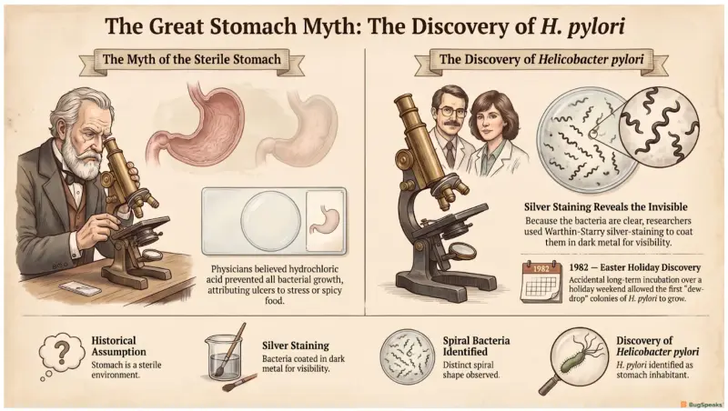
For thousands of years, people suffered from terrible stomach aches and burning pains called ulcers. Doctors long ago believed that no tiny pathogen could ever live inside the human stomach because our digestive juices are full of burning hydrochloric acid. Because this hidden bacterium is completely clear and cannot be seen under normal microscopes, it remained a total ghost to science. People assumed that painful stomach sores were just caused by stress, eating spicy foods, or cold weather.
Everything changed when two brave scientists decided to look much closer at human tissue samples using a special silver-staining technique. This brilliant method coated the invisible, clear bacteria in dark metal, making them pop out like shadows under the lens. In 1982, these researchers accidentally left their culture plates in an incubator over an extended Easter holiday weekend. When they returned, they discovered tiny, shiny, dew-drop colonies of a brand-new, spiral-shaped creature that had been hiding in plain sight.
The scientists initially named this mysterious, twisting microorganism Campylobacter pyloridis because it looked like other curved germs. However, after analyzing its unique 16S rRNA genetic code, they realized it belonged to a completely separate family tree. In 1989, they officially renamed it Helicobacter pylori, combining the Greek word for spiral with the word for rod. One scientist even drank a cloudy beaker of the live germs to prove they caused stomach sickness!
Why Does This Fragile Parasite Need a Human Body to Stay Alive?
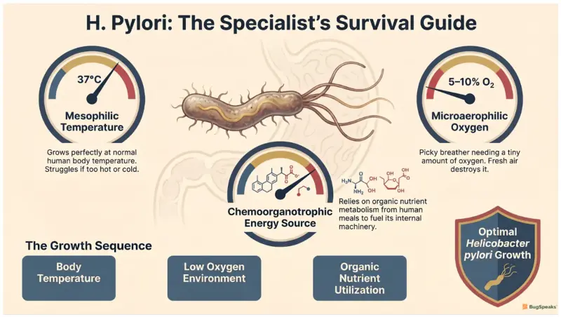
This special microbe is highly customized to live inside the warm, cozy walls of our digestive system. It is classified as a mesophilic organism, which means it thrives best at a normal human body temperature of 37°C. If it gets too cold or too hot, it cannot multiply properly and struggles to survive. It is also quite picky about its breathing conditions, demanding a very specific microaerophilic atmosphere. This means it needs just a tiny bit of oxygen to breathe, but too much fresh air will actually destroy it.
To keep its internal machinery running, this germ relies on a chemoorganotrophic lifestyle, meaning it breaks down organic chemicals for food. It absorbs special amino acids and sugars from our meals to generate its energy. While it loves the human body, it can transform into an inactive, round ball when stressed. This clever shape-shifting trick allows it to sleep in external reservoirs like municipal drinking water, raw milk, and river systems. This state helps it wait patiently until it can find a brand-new human host.
Our little spiral friend is not always a bad guy, as it has co-evolved with humans for 100,000 years. Having this microbe around can actually train our immune systems and protect kids from developing severe allergies or asthma. It balances out our body chemistry, but when things go wrong, it can cause major trouble.
How Does This Living Corkscrew Drill Through Our Internal Defenses?
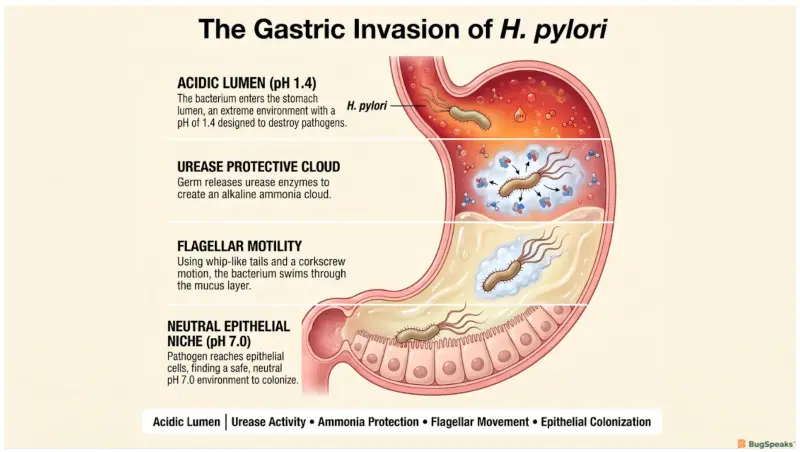
The stomach is a dangerous place, featuring a highly acidic environment that easily destroys most normal bacteria. To survive this harsh puddle, the microbe uses a superpower called urease activity to build a protective shield. This special enzyme breaks down a chemical called urea into ammonia and carbon dioxide. The alkaline ammonia forms a cozy, basic cloud around the germ, neutralizing the burning acid nearby. This incredible trick protects the outer membrane of the cell wall from being melted by our digestive juices.
Once protected, the bacterium uses its spiral shape and long, whip-like tails to swim forward. It detects chemical clues to steer away from acid and head straight toward the safe stomach wall. The ammonia cloud liquefies the thick, jello-like mucus layer protecting our stomach lining, turning it into a runny fluid. This allows the germ to rotate like a corkscrew and drill deep into the tissue. The basic steps of this invasion process follow a very specific path:
[Neutralize Acid] ──► [Liquefy Mucus] ──► [Drill Tissue] ──► [Inject Toxins]
Once inside the deep tissue, the bacterium forms a tough, sticky community known as a biofilm matrix. This gooey fortress shields the microbes from moving fluids, immune system cells, and incoming medical treatments. Safely tucked away inside this slime capsule, the pathogen can release dangerous toxins that irritate our cells. These toxic proteins damage the stomach lining, causing painful, inflamed sores that make people feel very sick.
How Did Fighting This Microbe Launch the Era of Magic Bullets and Fever Cures?
Long before modern antibiotics were invented, doctors discovered that applying extreme heat could sometimes stop certain long-term infections. Because this bacterium lacks special heat-shock protection genes, it cannot handle high temperatures very well. Early researchers noticed that high fevers would accidentally cook the fragile germs right inside the human body. This crazy observation helped scientists realize that every single microbe has a unique weakness that can be targeted. It launched a frantic search for specific chemical weapons that could kill germs without harming patients.
When modern medicine arrived, doctors created a standard treatment plan combining heavy acid-blockers with strong antibiotic medicines. Unfortunately, this clever germ started changing its own DNA to fight back against our strongest standard drugs. It developed a powerful antimicrobial resistance, allowing it to survive treatments that used to wipe it out completely. When it survives these drugs, it creates a stubborn, long-lasting condition called a refractory infection. This means the normal medical tools fail, forcing scientists to search for brand-new ways to beat them.
To solve this problem, doctors are now using advanced probiotics to help out. These friendly, helpful bacteria are packed into foods like yogurt to support our internal ecosystem. When we swallow these good microbes, they rush into the stomach and pick a fight with the bad germs. They crowd out the spiral invaders, break down their sticky biofilm fortresses, and restore peace to our tummies.
How Are Modern Genetic Blueprints Helping Us Create the Ultimate Shield?
Today, scientists are using incredible genetic tools to track this ancient organism across the planet. By studying its unique DNA, researchers can use the microbe as a phylogeographic marker to trace human history. Because this germ is passed down through families, its mutations match the ancient migration routes of early humans. Scientists even extracted this bacterium from the stomach of a 5,300-year-old frozen mummy named Ötzi the Iceman! This ancient genetic blueprint proved that early Europeans suffered from stomach aches thousands of years ago.
In high-tech laboratories, bioengineers are completely reversing the germ's machinery to help save lives through recombinant DNA technology. Instead of letting the bacterium cause harm, scientists cut out its bad genes and insert helpful instructions. They use the germ's highly active promoter systems to create useful therapeutic proteins directly inside living tissues. This allows the modified bacteria to act like microscopic factories, delivering helpful medicines to the body.
Scientists are also working hard to build a protective shield using a modern oral vaccine. These advanced treatments train our bodies to recognize specific pieces of the germ, like its outer coat proteins. When a child takes the vaccine, their immune system creates specialized defensive proteins to fight off future invasions. By teaching our bodies to recognize the spiral shape early, we can stop the pathogen before it ever starts.
Taxonomic Classification
Microbe Profile
Shape- Helical, spiral, or S-shaped curved rod; can turn into a spherical coccoid shape under stress.
Gram Stain Nature- Gram-negative cell wall structure with an outer membrane containing lipopolysaccharides.
Spore-forming- Asporogenous (does not form endospores).
Biofilm formation- Forms structured biofilms at air/liquid interfaces and on stomach linings using a complex sugar matrix regulated by the luxS and cagE genes.
Oxygen requirements- Microaerophilic
Optimal Temperature- Mesophilic (grows best at 37°C).
Optimal pH- Replicates best at a neutral pH of 6.0 to 8.5, but survives acid down to pH 4.0 using its active urease buffer.
Nutrient Usage- Chemoorganotrophic respiratory metabolism; consumes glucose and essential amino acids (like arginine, leucine, and valine).
Fun Facts
The Birthday Bacilli: Dr. Robin Warren first spotted the beautiful, glowing blue lines of these hidden spiral bacteria on a human tissue slide on June 11, 1979, which happened to be his 42nd birthday!
The Heroic Cocktail: To prove to the world that this germ was the real culprit behind painful stomach ulcers, Australian scientist Barry Marshall mixed up a warm broth of cultured bacteria and drank it himself, eventually winning a Nobel Prize for his bravery.
A Heavyweight Enzyme: The protective urease enzyme made by this microbe is an absolute giant with a molecular weight of 580 kDa, and the cell makes so much of it that it takes up a large part of the bacterium's entire protein weight!
Reference
Öztekin, M., Yılmaz, B., Ağagündüz, D., & Capasso, R. (2021). Overview of Helicobacter pylori Infection: Clinical Features, Treatment, and Nutritional Aspects. Diseases (Basel, Switzerland), 9(4), 66. https://doi.org/10.3390/diseases9040066
Cellini L. (2014). Helicobacter pylori: a chameleon-like approach to life. World journal of gastroenterology, 20(19), 5575–5582. https://doi.org/10.3748/wjg.v20.i19.5575
Yang, J. C., Lu, C. W., & Lin, C. J. (2014). Treatment of Helicobacter pylori infection: current status and future concepts. World journal of gastroenterology, 20(18), 5283–5293. https://doi.org/10.3748/wjg.v20.i18.5283
Lee, W. C., Goh, K. L., Loke, M. F., & Vadivelu, J. (2017). Elucidation of the Metabolic Network of Helicobacter pylori J99 and Malaysian Clinical Strains by Phenotype Microarray. Helicobacter, 22(1), e12321. https://doi.org/10.1111/hel.12321
Kusters, J. G., van Vliet, A. H., & Kuipers, E. J. (2006). Pathogenesis of Helicobacter pylori infection. Clinical microbiology reviews, 19(3), 449–490. https://doi.org/10.1128/CMR.00054-05
Graham, D. Y., & Dore, M. P. (2016). Helicobacter pylori therapy: a paradigm shift. Expert review of anti-infective therapy, 14(6), 577–585. https://doi.org/10.1080/14787210.2016.1178065
Yamaoka Y. (2009). Helicobacter pylori typing as a tool for tracking human migration. Clinical microbiology and infection : the official publication of the European Society of Clinical Microbiology and Infectious Diseases, 15(9), 829–834. https://doi.org/10.1111/j.1469-0691.2009.02967.x
Moss, S. F., Shah, S. C., Tan, M. C., & El-Serag, H. B. (2024). Evolving Concepts in Helicobacter pylori Management. Gastroenterology, 166(2), 267–283. https://doi.org/10.1053/j.gastro.2023.09.047
Suzuki, R., Saitou, N., Matsuari, O., Shiota, S., Matsumoto, T., Akada, J., Kinjo, N., Kinjo, F., Teruya, K., Shimoji, M., Shiroma, A., Kato, M., Satou, K., Hirano, T., Asaka, M., Kryukov, K., Moodley, Y., & Yamaoka, Y. (2022). Helicobacter pylori genomes reveal Paleolithic human migration to the east end of Asia. iScience, 25(7), 104477. https://doi.org/10.1016/j.isci.2022.104477