# Mouth Breathing vs Nose Breathing: Impact on Oral and Gut Health
Author: Neha Rao
Author URL: https://www.bugspeaks.com/blog/author/neha-rao
Published: 2026-09-13
Category: Microbiome and Lifestyle
Category URL: https://www.bugspeaks.com/blog/category/microbiome-and-lifestyle
Meta Title: Mouth Breathing vs Nose Breathing: Oral Health | BugSpeaks
Meta Description: Learn how mouth breathing affects oral health and gut health by drying saliva, shifting oral pH, and promoting microbial imbalance. Discover the connection!
Tags: Gut Health, Mouth Breathing, Nose Breathing
Tag URLs: Gut Health (https://www.bugspeaks.com/blog/tag/gut-health), Mouth Breathing (https://www.bugspeaks.com/blog/tag/mouth-breathing), Nose Breathing (https://www.bugspeaks.com/blog/tag/nose-breathing)
URL: https://www.bugspeaks.com/blog/mouth-nose-breathing-gut-health
## **Why does breat** hing through your mouth during sleep change the oral environment?
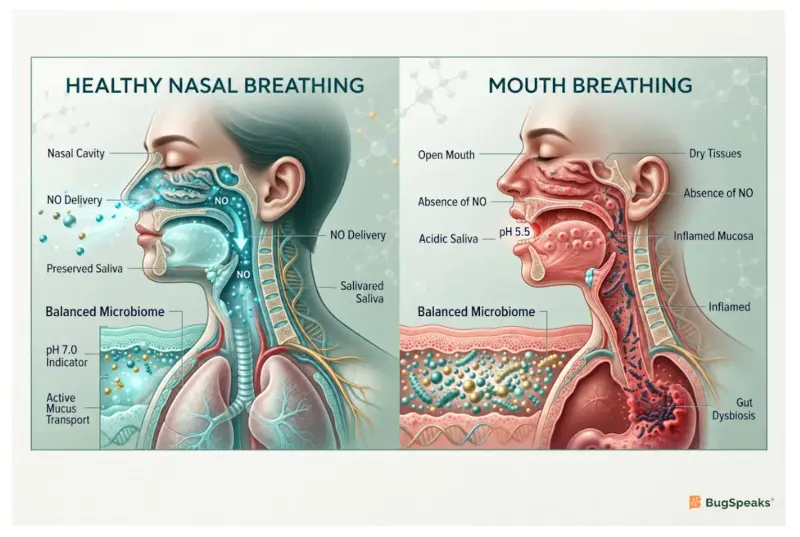
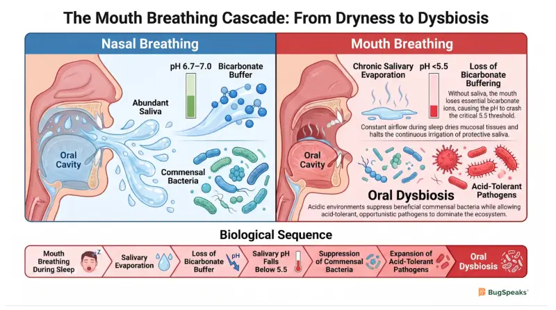
Mouth breathing during [sleep](https://www.bugspeaks.com/blog/sleep-circadianrhythm-guthealth-gutmicrobiome) completely alters the oral environment by drying out mucosal tissues, reducing salivary flow, and lowering the potential of hydrogen (pH) to an acidic range. In a [healthy](https://www.bugspeaks.com/blog/high-heels-digestive-flow-gut-health) state, breathing through your nose keeps your mouth closed, allowing saliva to coat your teeth and gums continuously. This natural fluid serves as the primary transport and balancing medium of the [oral-gut](https://www.bugspeaks.com/blog/hiit-yoga-gut-transit-time) gateway, keeping tissues moist. When you breathe through your mouth at night, the constant airflow evaporates this moisture, leading to chronic dryness. This stops the steady irrigation of the mouth, which is essential for biological health and local homeostasis [Fan et al. (2020)](https://doi.org/10.3389/fmicb.2020.575550).
To clinically diagnose mouth breathing in children, examiners perform several specialized clinical tests that evaluate nasal patency and airflow. These include the water retention test, where a child must retain water in their mouth for a set period, the single nose test, the mirror test to check for condensation, and the cotton batting test to observe physical movement. In children identified as mouth breathers, a thorough laryngological examination often reveals underlying chronic conditions like rhinitis, nasosinusitis, or adenoidal hypertrophy. These conditions reduce the openness of the nasal airways, forcing the child to rely on mouth breathing, which initiates a pathological cascade across the oral mucosa [Fan et al. (2020)](https://doi.org/10.3389/fmicb.2020.575550).
Without saliva, the oral cavity loses its primary protective buffer, causing a rapid drop in salivary pH that damages tissues. Saliva naturally contains bicarbonate and phosphate ions that neutralize the acids produced by oral bacteria such as Streptococcus mutans, Streptococcus sobrinus, and Lactobacillus species during their metabolic processes [Tansel and Levinthal (2023)](https://doi.org/10.14309/ctg.0000000000000567). When mouth breathing dries out this fluid, the protective buffer is lost. The protective buffer is a combination of bicarbonate and phosphate ions dissolved in saliva. These natural ions bind to and neutralize bacterial acids, keeping your mouth at a healthy, non-corrosive pH. When the intraoral environment becomes increasingly acidic, it often drops below the critical salivary pH threshold of 5.5 [Fan et al. (2020)](https://doi.org/10.3389/fmicb.2020.575550). This acidic shift completely alters the oral ecosystem, creating a selective environment where beneficial, acid-sensitive bacteria such as Streptococcus sanguinis, Streptococcus gordonii, Streptococcus mitis, Rothia species, and Olsenella species
are suppressed while acid-tolerant species thrive, initiating a state of localized oral dysbiosis [Fan et al. (2020)](https://doi.org/10.3389/fmicb.2020.575550).
**Mouth breathing**\- Breathing through your mouth instead of your nose, which dries out saliva.
**Mouth breathers**\- People who breathe through their mouth, often due to a blocked nose.
**Saliva**\- The natural fluid in your mouth that washes away food and neutralizes acid.
**Salivary pH**\- The scale that measures the acid levels in your saliva, normally close to neutral.
**Homeostasis**\- A balanced, healthy state of your body where everything functions normally.
**Oral dysbiosis**\- An imbalance of the microbes in your mouth, where bad bacteria outnumber good ones.
**Nasal patency**\- How clear and open your nasal passages are for air to flow smoothly.
**Water retention test**\- A simple test where you hold water in your mouth for a few minutes to see if you can breathe through your nose.
**Single nose test**\- A clinical test where one nostril is blocked at a time to check airflow through each side of your nose.
**Mirror test**\- A test that measures condensation on a cool mirror placed under your nose to check for nasal airflow.
**Laryngological examination**\- A physical checkup by an ear, nose, and throat doctor to inspect your throat and upper airways.
**Nasosinusitis**\- Swelling and irritation of the tissues lining both your nose and your sinus cavities.
**Adenoidal hypertrophy**\- Enlarged glands at the back of your nasal cavity that can block normal nose breathing.
**Rhinitis**\- Inflammation of the inner lining of your nose, often causing stuffiness or a runny nose.
## **How** does a dry mouth disrupt the biological defenses of the oral-gut gateway?
A dry mouth disrupts the biological defenses of the oral-gut gateway by reducing the levels of protective immune proteins and increasing markers of cellular oxidative stress. Under normal conditions, saliva contains a suite of defensive proteins, including secretory immunoglobulin A (sIgA), lysozymes, and lactoferrins. These proteins actively inhibit the growth of opportunistic pathogens such as Acinetobacter calcoaceticus, Neisseria species, Streptococcus pneumoniae, Stenotrophomonas species, Fusobacterium nucleatum, and Porphyromonas gingivalis, and prevent them from adhering to the oral mucosa [Mantis et al. (2011)](https://doi.org/10.1038/mi.2011.41). When mouth breathing dries out the oral cavity, the production and concentration of these natural protective proteins decrease significantly, leaving the mucosal surface vulnerable to colonization by harmful pathogens, which compromises the entry point of the digestive tract [Fan et al. (2020)](https://doi.org/10.3389/fmicb.2020.575550).
This reduction in mucosal immunity is accompanied by a significant increase in salivary markers of cell damage and oxidative stress. Scientific studies have demonstrated that mouth-breathing children exhibit a significant upregulation of oxidative-stress-related salivary proteins, specifically lactoylglutathione lyase (GLO1) and peroxiredoxin-5 [Fan et al. (2020)](https://doi.org/10.3389/fmicb.2020.575550). This specialized lactoylglutathione lyase is an enzyme involved in the detoxification of methylglyoxal, which is a toxic byproduct of cellular glycolysis. The elevated levels of lactoylglutathione lyase indicate that the cells lining the dry oral cavity are under severe metabolic stress, struggling to neutralize toxic compounds in the absence of adequate saliva [Fan et al. (2020)](https://doi.org/10.3389/fmicb.2020.575550).
Similarly, peroxiredoxin-5 (PRDX5) acts as a protective antioxidant enzyme by chemically breaking down harmful reactive hydrogen peroxide into harmless water and oxygen before it can damage cell membranes and chemical oxidative attacks. Other proteins, such as integrin alpha-M and proteasome subunit alpha type-1, provide critical protective functions in the oral cavity but are downregulated in mouth breathers, which compromises cellular integrity [Fan et al. (2020)](https://doi.org/10.3389/fmicb.2020.575550). These protein alterations indicate that mouth breathing not only depletes the mouth's physical defenses but also causes a metabolic shift that supports a pro-inflammatory microenvironment. This weakens the mucosal barrier, allowing pathogens to multiply and prepare for pathological translocation down the digestive tract [Glavina et al. (2026)](https://doi.org/10.3390/life16020294).
**Defensive Component / Marker**
**Normal Function in Oral-Gut Gateway**
**Alteration Status from Mouth Breathing**
**Downstream Health Impact on Your Body**
Secretory immunoglobulin A
Glues harmful bacteria together to wash them away safely
Significantly Decreased
Pathogens adhere easily to mucosal tissues [Mantis et al. (2011)](https://doi.org/10.1038/mi.2011.41)
Oxidative stress Markers
Protects cells from damage and chemical attacks
Significantly Increased
Triggers cell irritation and tissue damage [Fan et al. (2020)](https://doi.org/10.3389/fmicb.2020.575550)
Lactoylglutathione lyase
Detoxifies harmful glycolytic stress byproducts
Significantly Increased
Signals elevated cellular metabolic stress [Fan et al. (2020)](https://doi.org/10.3389/fmicb.2020.575550)
Peroxiredoxin-5
Antioxidant enzyme protecting cells from peroxides
Significantly Increased
Marks cellular defense against oxidative damage [Fan et al. (2020)](https://doi.org/10.3389/fmicb.2020.575550)
**Secretory immunoglobulin A**\- A protective protein antibody in saliva that traps bacteria.
**Oxidative stress**\- Cell damage caused by harmful chemicals when your body is under stress.
**Lactoylglutathione lyase**\- A special enzyme that cleans up toxic chemicals inside stressed cells.
**Peroxiredoxin-5**\- A protective antioxidant enzyme that shields cells from chemical damage.
**Lysozymes**\- Natural protective enzymes in saliva that break down bacterial walls to destroy germs.
**Lactoferrins**\- Protective proteins in saliva that bind up iron to starve bacteria of the nutrients they need to grow.
**Methylglyoxal**\- A toxic chemical byproduct formed inside cells during energy production that causes cellular stress if not cleared out.
**Integrin alpha-M**\- A protein on white blood cells that helps them stick to tissues so they can move to sites of infection.
**Proteasome subunit alpha type-1**\- A component of the cell's cleanup system that breaks down old or damaged proteins to keep cells healthy.
## **H** ow do oral bacteria migrate through the digestive tract to reach the gut?
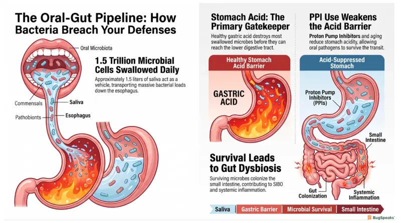
Oral bacteria migrate to the gut through the swallowing of saliva, which carries billions of microbial cells past the gastric acid barrier of the stomach into the lower digestive tract. The mouth and the gut are connected along a single physical axis, translocating oral microbes in a continuous process. Every day, a person swallows about 1.5 liters of saliva, which acts as a vehicle, transporting an estimated 1.5 trillion biological cells [Kamal et al. (2026)](https://doi.org/10.3389/fcimb.2026.1769355). Under normal circumstances, the highly acidic stomach serves as the first major chemical barrier, destroying most swallowed microbes before they can reach the intestines [Kamal et al. (2026)](https://doi.org/10.3389/fcimb.2026.1769355).
However, when oral dysbiosis occurs, the sheer volume and altered nature of the migrating bacteria can overwhelm the stomach's defenses. If a person has reduced gastric acid from aging, stress, or medications like proton pump inhibitors (PPIs), the survival rate of swallowed oral bacteria increases [Kamal et al. (2026)](https://doi.org/10.3389/fcimb.2026.1769355). Under these conditions, species that would normally be destroyed by stomach acid survive the transit and enter the small intestine [Tansel and Levinthal (2023)](https://doi.org/10.14309/ctg.0000000000000567). This survival is further enhanced because many oral bacteria, such as Fusobacterium nucleatum, Porphyromonas gingivalis, Streptococcus mutans, and Granulicatella adiacens form dense, protective biofilms that shield individual cells from acid exposure, allowing them to pass through the stomach intact [Kamal et al. (2026)](https://doi.org/10.3389/fcimb.2026.1769355).
Once they survive the stomach transit, these oral microbes enter the distinct ecosystem of the gut microbiome. If this influx leads to significant colonization of the small intestine, it can contribute to a condition known as small intestinal bacterial overgrowth (SIBO) [Tansel and Levinthal (2023)](https://doi.org/10.14309/ctg.0000000000000567). To evaluate this overgrowth non-invasively, clinicians use hydrogen-methane breath testing, which measures exhaled gases produced by microbial fermentation of carbohydrates [Tansel and Levinthal (2023)](https://doi.org/10.14309/ctg.0000000000000567). A rise of 20 parts per million in hydrogen gas from baseline within 90 minutes is diagnostic of SIBO, highlighting how upstream oral health directly impacts gut stability [Tansel and Levinthal (2023)](https://doi.org/10.14309/ctg.0000000000000567).
**Gastric acid**\- The powerful acid inside your stomach that kills swallowed bacteria.
**Proton pump inhibitors**\- Medications that reduce stomach acid, which can let bacteria survive the journey.
**Small intestinal bacterial overgrowth**\- A condition where too many bacteria colonize the small intestine, causing bloating.
**Hydrogen-methane breath testing**\- A non-invasive test where you breathe into a tube to measure gases made by bacteria in your gut.
## What happens when opportunistic oral pathogens invade the gut microbiome?
When opportunistic oral pathogens invade the gut microbiome, they disrupt the local ecosystem's balance, compromise the intestinal barrier, and trigger a low-grade systemic inflammatory cascade. Once oral pathogens like Fusobacterium nucleatum or Porphyromonas gingivalis translocate to the gut, they use specialized adhesion proteins to bind to intestinal epithelial cells [Kamal et al. (2026)](https://doi.org/10.3389/fcimb.2026.1769355). For instance, Fusobacterium nucleatum utilizes its FadA adhesin to attach to epithelial cells, disrupting the tight junctions that hold the intestinal barrier together [Kamal et al. (2026)](https://doi.org/10.3389/fcimb.2026.1769355). This structural damage increases the permeability of the gut lining, allowing harmful substances to leak into the bloodstream [Glavina et al. (2026)](https://doi.org/10.3390/life16020294).
The role of mucosal immunity is critical in preventing this invasion. In healthy tissues, secretory immunoglobulin A acts as a protective shield, binding to pathogens and keeping them in the lumen [Mantis et al. (2011)](https://doi.org/10.1038/mi.2011.41). However, when a localized secretory IgA deficiency occurs on the mucosal surface of the small airways or the intestines, colonizing bacteria are able to cross the epithelial barrier [Polosukhin et al. (2017)](https://doi.org/10.1164/rccm.201604-0759OC). This bacterial invasion activates nuclear factor-kappa B signaling in the epithelial cells [Polosukhin et al. (2017)](https://doi.org/10.1164/rccm.201604-0759OC). The activation of this pathway triggers the release of pro-inflammatory cytokines, driving persistent local inflammation and tissue remodeling [Polosukhin et al. (2017)](https://doi.org/10.1164/rccm.201604-0759OC).
This breach of the barrier enables bacterial endotoxins, such as lipopolysaccharide (LPS), to enter the systemic circulation [Adil et al. (2025)](https://doi.org/10.3390/microorganisms13040814). Lipopolysaccharide is a major component of the cell wall of Gram-negative bacteria and serves as a potent trigger for the host immune system [Adil et al. (2025)](https://doi.org/10.3390/microorganisms13040814). When lipopolysaccharide leaks into the bloodstream, it binds to Toll-like receptors on immune cells, initiating a cascade that releases pro-inflammatory cytokines like interleukin-6 and tumor necrosis factor-alpha [Adil et al. (2025)](https://doi.org/10.3390/microorganisms13040814). This process leads to metabolic endotoxemia and chronic low-grade inflammation, which can affect metabolic organs, insulin sensitivity, and overall systemic health [Adil et al. (2025)](https://doi.org/10.3390/microorganisms13040814).
**Intestinal barrier**\- The protective wall lining your gut that keeps bad materials out of your blood.
**Fusobacterium nucleatum**\- A common mouth bacterium that can travel down and irritate your intestines.
**Porphyromonas gingivalis**\- A periodontal pathogen that triggers systemic inflammation when it spreads.
**Lipopolysaccharide**\- A toxic component of bacterial walls that causes a massive immune response.
**FadA adhesin**\- A protein hook on the surface of _Fusobacterium nucleatum_ that helps it stick tightly to gut wall cells.
**Nuclear factor-kappa B**\- A control protein inside cells that switches on inflammatory genes when danger or bacteria are detected.
**Pro-inflammatory cytokines**\- Chemical signals released by immune cells that tell the body to create swelling and inflammation.
**Gram-negative bacteria**\- A group of bacteria with a double-layered outer wall containing toxins that can trigger immune responses.
**Interleukin-6**\- An immune messenger protein that signals the body to cause swelling and inflammation during an infection.
**Tumor necrosis factor-alpha**\- A powerful inflammatory protein released by immune cells to alert the body to tissue damage or invaders.
**Endotoxemia**\- A condition where toxic bacterial wall pieces leak out of the gut and travel into your bloodstream.
## How does nose breathing protect the entire oral-gut pathway from microbial imbalance?
Nose breathing protects the entire oral-gut pathway by maintaining proper moisture levels in the mouth and facilitating the continuous release of antimicrobial airway gases. When you breathe through your nose, your mouth remains closed, preserving saliva and allowing it to perform its essential buffering and protective functions [Fan et al. (2020)](https://doi.org/10.3389/fmicb.2020.575550). This simple act keeps the oral salivary pH stable and ensures that natural defensive proteins remain active. The stable, healthy oral salivary pH range is 6.5 to 7.0. Furthermore, nose breathing allows the paranasal sinuses to produce and release nitric oxide (NO), a crucial gas with potent antimicrobial effects [Workman et al. (2017)](https://doi.org/10.1002/alr.21966), which keeps the oral ecosystem healthy and balanced by selectively destroying harmful pathogens (like Pseudomonas aeruginosa and Candida albicans) while leaving friendly commensal bacteria (like Staphylococcus epidermidis) unharmed, while simultaneously speeding up mucociliary clearance to sweep away trapped germs.
Nitric oxide serves as a vital component of the host's innate immune defense, exerting concentration-dependent bacteriostatic and bactericidal effects on invading pathogens [Workman et al. (2017)](https://doi.org/10.1002/alr.21966). Scientific research has shown that airway pathogens have varying susceptibilities to this gas at physiologic concentrations [Workman et al. (2017)](https://doi.org/10.1002/alr.21966). While common commensal bacteria like Staphylococcus epidermidis exhibit higher resistance to nitric oxide, allowing them to remain as healthy residents of the airway, virulent pathogens like Pseudomonas aeruginosa and fungi like Candida albicans are highly sensitive and are easily controlled [Workman et al. (2017)](https://doi.org/10.1002/alr.21966). This selective antimicrobial action helps maintain the balance of the healthy respiratory and oral microbiomes [Workman et al. (2017)](https://doi.org/10.1002/alr.21966).
In addition to its direct antimicrobial effects, inhaled nitric oxide stimulates ciliary beating, which accelerates mucociliary clearance (MCC) and helps transport trapped pathogens to the oropharynx, where they are cleared safely [Workman et al. (2017)](https://doi.org/10.1002/alr.21966). By keeping the mouth closed and ensuring a steady flow of nitric oxide, nose breathing prevents the dry, acidic conditions that lead to oral dysbiosis [Workman et al. (2017)](https://doi.org/10.1002/alr.21966). This protection stabilizes the first node of the oral-gut gateway, preventing the overgrowth of harmful bacteria and ensuring that the flow of microbes down into the lower gut remains balanced, healthy, and non-pathogenic [Adil et al. (2025)](https://doi.org/10.3390/microorganisms13040814).
**Physiological Parameter**
**Nose Breathing Pathway**
**Mouth Breathing Pathway**
**Primary Biological Consequence**
Oral Surface Moisture
Preserved by closed mouth
Dried out by constant airflow
Salivary defense enzymes remain functional or get evaporated
Local Potential of Hydrogen
Balanced (neutral pH 6.5 - 7.0)
Acidic (critical pH below 5.5)
Prevents or promotes cariogenic pathogen overgrowth
Nitric oxide Delivery
Continuously inhaled from paranasal sinuses
Bypassed entirely
Selective killing of virulent pathogens [Workman et al. (2017)](https://doi.org/10.1002/alr.21966)
Mucociliary clearance
Stimulated by sinus gases
Slowed down due to lack of gases
Pathogens are cleared safely or colonize tissues [Workman et al. (2017)](https://doi.org/10.1002/alr.21966)
**Nitric oxide**\- A helpful gas made in your nose sinuses that selectively kills bad germs.
**Staphylococcus epidermidis**\- A friendly bacterium that lives safely in your nose and airways.
**Pseudomonas aeruginosa**\- A harmful bacterium that is highly sensitive to and killed by nitric oxide.
**Candida albicans**\- A common yeast that can cause infections when mouth defenses are weak.
**Mucociliary clearance**\- The sweeping movement of tiny hairs in your airways that clears out germs.
**Bacteriostatic**\- Stopping or slowing down the multiplication of bacteria without immediately killing them.
**Bactericidal effects**\- Actions or substances that directly kill bacterial cells.
**Oropharynx**\- The middle section of your throat located right behind your mouth.
Visualize the process- [https://youtu.be/NLKLgBclITA](https://youtu.be/NLKLgBclITA)
### Reference
Fan, C., Guo, L., Gu, H., Huo, Y., & Lin, H. (2020). Alterations in Oral-Nasal-Pharyngeal Microbiota and Salivary Proteins in Mouth-Breathing Children. _Frontiers in microbiology_, _11_, 575550\. [https://doi.org/10.3389/fmicb.2020.575550](https://doi.org/10.3389/fmicb.2020.575550)
Adil, N. A., Omo-Erigbe, C., Yadav, H., & Jain, S. (2025). The Oral-Gut Microbiome-Brain Axis in Cognition. _Microorganisms_, _13_(4), 814. [https://doi.org/10.3390/microorganisms13040814](https://doi.org/10.3390/microorganisms13040814)
Workman, A. D., Carey, R. M., Kohanski, M. A., Kennedy, D. W., Palmer, J. N., Adappa, N. D., & Cohen, N. A. (2017). Relative susceptibility of airway organisms to antimicrobial effects of nitric oxide. _International forum of allergy & rhinology_, _7_(8), 770–776. [https://doi.org/10.1002/alr.21966](https://doi.org/10.1002/alr.21966)
Kamal FZ, Lefter R, Ciobica A, Burlui V, Rammali S, Abdou A, Grigore M-N, Ionescu C, Tomita D-I and Gheban MD (2026) From gut to mouth: salivary signatures of carcinogenic microbiota. Front. Cell. Infect. Microbiol. 16:1769355. doi: 10.3389/fcimb.2026.1769355
Mantis, N. J., Rol, N., & Corthésy, B. (2011). Secretory IgA's complex roles in immunity and mucosal homeostasis in the gut. _Mucosal immunology_, _4_(6), 603–611. [https://doi.org/10.1038/mi.2011.41](https://doi.org/10.1038/mi.2011.41)
Polosukhin, V. V., Richmond, B. W., Du, R. H., Cates, J. M., Wu, P., Nian, H., Massion, P. P., Ware, L. B., Lee, J. W., Kononov, A. V., Lawson, W. E., & Blackwell, T. S. (2017). Secretory IgA Deficiency in Individual Small Airways Is Associated with Persistent Inflammation and Remodeling. _American journal of respiratory and critical care medicine_, _195_(8), 1010–1021. [https://doi.org/10.1164/rccm.201604-0759OC](https://doi.org/10.1164/rccm.201604-0759OC)
Xiao, J., Fiscella, K. A., & Gill, S. R. (2020). Oral microbiome: possible harbinger for children's health. _International journal of oral science_, _12_(1), 12. [https://doi.org/10.1038/s41368-020-0082-x](https://doi.org/10.1038/s41368-020-0082-x)
Tansel, A., & Levinthal, D. J. (2023). Understanding Our Tests: Hydrogen-Methane Breath Testing to Diagnose Small Intestinal Bacterial Overgrowth. _Clinical and translational gastroenterology_, _14_(4), e00567. [https://doi.org/10.14309/ctg.0000000000000567](https://doi.org/10.14309/ctg.0000000000000567)
Glavina, A., Martić, D., Perko, M. A., Mešin Delić, D., Tadin, A., Lešić, S., & Šupe-Domić, D. (2026). The Oral Microbiome and Systemic Health: Current Insights into the Mouth-Body Connection. _Life (Basel, Switzerland)_, _16_(2), 294. [https://doi.org/10.3390/life16020294](https://doi.org/10.3390/life16020294)
## FAQs
Q: Does nose breathing during the day help improve gut health?
A: Yes, nose breathing keeps the mouth closed, preserving saliva and stabilizing the oral microbiome during the day Fan et al. (2020). This prevents upstream oral dysbiosis, which ensures that only a balanced and non-pathogenic mix of microbes is translocated down to the gut microbiome Adil et al. (2025).
Q: How does mouth breathing affect the risk of tooth decay?
A: Mouth breathing increases the risk of tooth decay by evaporating saliva, which depletes the mouth's natural acid-neutralizing buffers. In this highly acidic, dry environment, beneficial commensal bacteria cannot survive, whereas acid-tolerant, cavity-causing pathogens multiply rapidly within the oral biofilm Fan et al. (2020).
Q: What is the relationship between proton pump inhibitors and bacterial translocation?
A: Proton pump inhibitors suppress the production of gastric acid in the stomach, which weakens the body's first major chemical barrier. This reduction in stomach acidity allows swallowed oral bacteria to survive the transit through the stomach, significantly increasing their survival and colonization within the lower digestive tract Tansel and Levinthal (2023).
Q: Why are some airway bacteria more sensitive to nitric oxide than others?
A: Different bacterial species possess distinct physiological metabolic pathways and enzymatic scavengers that influence their susceptibility to paranasal gases Workman et al. (2017). For example, virulent pathogens are highly sensitive to the concentration-dependent bacteriostatic and bactericidal effects of nitric oxide, whereas common commensal bacteria possess natural resistance mechanisms that allow them to persist safely Workman et al. (2017).
Q: How can someone easily check if they are breathing through their mouth at night?
A: Common signs of mouth breathing during sleep include waking up with a very dry mouth, having chapped lips, or snoring. In clinical settings, examiners can also perform simple tests like the water retention test or check for condensation on a mirror placed under the nose to evaluate nasal patency Fan et al. (2020).
---
This blog is powered by Superblog. Visit https://superblog.ai to know more.
---