Understanding Estrobolome: A Guide to Hormone Balance and Acne
What is the estrobolome and how does it manage your body's hormone levels?
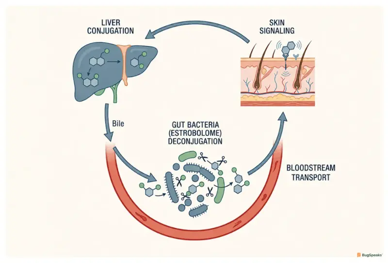
Inside your gut, a helpful bacterial crew called the estrobolome acts like a traffic controller, choosing exactly how much estrogen is allowed to re-enter your body and how much is safely cleared out as waste Lephart and Naftolin (2022). To understand this, imagine your body as a Hormone Exit & Re-Entry System that manages these hormone passengersLarnder et al. (2025). Active hormones like estrogen are departing molecules scheduled to leave once their work is finishedLarnder et al. (2025). Before these departing molecules can exit, they must pass a checkpoint that determines if they leave or receive a ticket to returnLephart and Naftolin (2022).
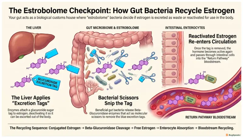
The journey of these departing molecules begins in your liver, which serves as the exit preparation site for hormonesLephart and Naftolin (2022). At this site, the liver modifies highly active estrogen to make it safe and water-soluble through a process called conjugationLephart and Naftolin (2022). This task is done by enzymes known as uridine diphosphate-glucuronosyltransferase (UGT) enzymes, which attach a chemical sugar tag to each hormoneLephart and Naftolin (2022). Once tagged, these inactive hormones travel into the intestine, which acts as the departure checkpoint, where they wait to either be excreted or prepared for re-entryLephart and Naftolin (2022).
Normally, this tagged package travels through the gut and exits the body via the waste, but the estrobolome can change this planLarnder et al. (2025). If the bacterial crew determines that the body needs more active hormones, they use an enzyme called beta-glucuronidase to perform deconjugationLephart and Naftolin (2022). This enzyme acts as a re-entry mechanism that snips the sugar tag right off the departing moleculeLarnder et al. (2025). Once the tag is removed, the hormone becomes active again, gets a return ticket, is reabsorbed, and enters the bloodstream return pathwayLephart and Naftolin (2022). This continuous loop is known as enterohepatic recirculationLephart and Naftolin (2022).
How do hormones travel through the Hormone Exit & Re-Entry System to influence your skin?
Hormones travel from your liver, pass through your gut checkpoint, and return via the bloodstream to interact directly with receptor proteins on your skin cells, which control skin cell growth, moisture, and inflammationLephart and Naftolin (2022). In this system, the bloodstream acts as the return pathway, transporting reactivated departing molecules from the gut checkpoint back to active duty throughout the bodyLephart and Naftolin (2022). When hormones are given return tickets at the gut checkpoint, they travel along this return pathway and arrive at the skinLephart and Naftolin (2022). This direct communication network linking our gut to our skin is called the gut-skin axisZhao et al. (2025).
To understand how these returning molecules affect your skin, we must look at the receptors waiting at the destinationLephart and Naftolin (2022). Your skin cells have specialized landing pads called estrogen receptors, which bind these hormonesLephart and Naftolin (2022). The most prominent landing pad is estrogen receptor beta, which binds estrogen to regulate key dermal processesLephart and Naftolin (2022). When the departing molecules are recycled via the enterohepatic recirculation loop and bind to these receptors, they stimulate collagen, promote cellular turnover, and maintain the skin barrierLephart and Naftolin (2022). This signaling keeps your skin thick and hydratedLephart and Naftolin (2022).
This hormonal path is further regulated by other enzymes at our gut checkpoint that manage different steroid pathwaysLarnder et al. (2025). For instance, enzymes called hydroxysteroid dehydrogenases (HSDs), including 3β-hydroxysteroid dehydrogenase and 17β-hydroxysteroid dehydrogenase, help interconvert different hormone precursors in gut bacteriaLarnder et al. (2025). These microbial workers can transform inactive precursors into active hormones right at the checkpoint, adding another layer of complexity to the volume of molecules entering the return pathwayLarnder et al. (2025). When these pathways cooperate smoothly, the systemic hormone pool remains stable, and the skin receives exactly what it needsLephart and Naftolin (2022).
Why does an imbalance in your gut checkpoint trigger stubborn acne?
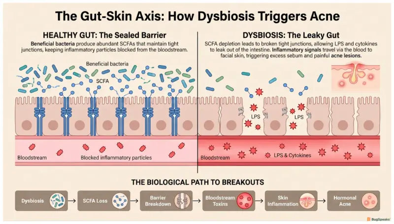
An imbalance in your gut checkpoint, known as dysbiosis, triggers skin inflammation and acne because it alters the amount of recycled hormones returning to your blood while disrupting the protective molecules that keep your skin barrier strongZhao et al. (2025). When your gut checkpoint experiences dysbiosis, the specialized bacterial crew becomes disorganized, allowing opportunistic pathogens to outcompete beneficial, protective microbesZhao et al. (2025). This chaos alters the production of beta-glucuronidase enzymes, leading to erratic deconjugation and causing wild swings in the volume of hormones entering the return pathwayLarnder et al. (2025). These fluctuations destabilize skin oil glandsZhao et al. (2025).
The breakdown of the checkpoint also directly damages the gut barrier itself, which relies on beneficial microbes to stay intactZhao et al. (2025). Healthy checkpoint bacteria ferment dietary fiber to produce beneficial molecules called short-chain fatty acids (SCFAs), such as butyrate, which keep the checkpoint walls tightly sealedZhao et al. (2025). In a state of dysbiosis, the production of these protective short-chain fatty acids drops significantly, causing the intestinal lining to become leaky and permeableZhao et al. (2025). This leakiness allows inflammatory bacterial fragments and toxic metabolites to escape the checkpoint and slip into the return pathwayZhao et al. (2025).
Once in the bloodstream, these inflammatory signals travel straight to the skin, where they trigger systemic immune activation and cause severe, red, and painful breakoutsZhao et al. (2025). This systemic inflammation works hand-in-hand with the hormonal disruptions caused by the malfunctioning estrobolomeLephart and Naftolin (2022). Under this dual stress, the sebaceous glands are stimulated to produce thick, excess sebum, which clogs pores and creates a perfect environment where acne-causing bacteria thriveZhao et al. (2025). The result is acne vulgaris (AV), a chronic inflammatory skin disease that is extremely difficult to treat with simple topical creamsZhao et al. (2025).
What is the connection between testosterone, the gut microbiota, and skin health?
Elevated levels of testosterone and other male hormones, a condition known as hyperandrogenism, are closely linked to a less diverse gut microbiome and increased sebum production, which directly promotes severe, inflammatory acneLi et al. (2024). In women, this hormonal excess is a defining feature of polycystic ovary syndrome (PCOS), a highly prevalent metabolic and reproductive disorder that affects millions of women worldwideLi et al. (2024). When active androgens like testosterone flood your entire body, they act as powerful signals that fundamentally alter the composition, diversity, and efficiency of your intestinal checkpoint crewLi et al. (2024). This disruption can lead to chronic skin inflammationZhao et al. (2025).
A landmark study byLi et al. (2024) revealed that women with hyperandrogenic polycystic ovary syndrome (PCOS) exhibit a dramatic decrease in the richness and diversity of their gut microbiota compared to healthy controls. At their intestinal checkpoint, beneficial, fiber-fermenting bacterial families like Bacteroidaceae and Oscillospiraceae are severely depleted, which reduces the production of protective anti-inflammatory metabolitesLi et al. (2024). Instead, their gut checkpoint is heavily dominated by pro-inflammatory bacterial families such as Enterobacteriaceae and Streptococcaceae, which promote local and systemic inflammation, weakening the checkpoint walls and triggering systemic flares and inflammatory acneLi et al. (2024). This makes their skin highly vulnerable to breakoutsZhao et al. (2025).
The study also identified key steroid hormones such as corticosterone, 5β-androsterone, and deoxycorticosterone that are significantly upregulated in the blood of patients, serving as potential biomarkers of this disrupted stateLi et al. (2024). These elevated cortical and androgenic hormones drive metabolic complications, weight gain, and systemic stress responsesLi et al. (2024). Interestingly, the researchers found that levels of testosterone negatively correlate with vitamin D3, suggesting that a deficiency in this nutrient acts as a major susceptibility factor that further destabilizes the gut-skin axis, letting inflammatory signals pass unchecked to create stubborn breakoutsLi et al. (2024). Supplementing vitamin D3 may restore this balanceLi et al. (2024).
How can we support our hormone exit and re-entry checkpoint through diet and lifestyle?
We can support our hormone exit and re-entry checkpoint by consuming a fiber-rich diet that feeds beneficial bacteria, using plant-based compounds to balance hormone receptors, and managing daily stress and sleep to stabilize gut-skin communicationLephart and Naftolin (2022). Shifting our diet is one of the most immediate and powerful ways to alter the composition and performance of our gut checkpoint crewLephart and Naftolin (2022). Diets rich in animal proteins and saturated fats are associated with higher fecal beta-glucuronidase activity, which leads to excessive hormone re-entry and potential hormonal imbalancesLephart and Naftolin (2022). Fiber significantly decreases its activityLephart and Naftolin (2022).
In addition to fiber, we can introduce protective plant-based compounds known as phytoestrogens, which are found in abundance in foods like soybeans, flaxseeds, and berriesLarnder et al. (2025). When these plant molecules reach the gut checkpoint, specialized microbial enzymes called beta-glucosidases perform a critical transformation, converting inactive plant glycosides into highly active, free-floating moleculesLarnder et al. (2025). These active compounds act as selective estrogen receptor modulators (SERMs), which can bind to estrogen receptors on skin cells and act as natural helpersLephart and Naftolin (2022). They help stabilize estrogen signaling, reduce skin inflammation, and boost hydrationLephart and Naftolin (2022).
Beyond diet, our daily habits and behaviors play a massive role in regulating the gut-skin communication network, helping to stabilize our biological systemsLephart and Naftolin (2022). Sleep deprivation, physical inactivity, and chronic stress directly trigger the release of stress hormones like cortisol, which damages the physical integrity of the gut barrier and causes a rapid shift toward dysbiosisLephart and Naftolin (2022). Practicing stress-reduction techniques and getting quality sleep help keep the gut lining intact, reduce the spillover of inflammatory markers, and ensure that the entire system can operate with maximum precision, efficiency, and long-term stabilityZhao et al. (2025).
-Varsha V
Visualize the process- https://youtu.be/9NIHe9u8KHU
Reference
Larnder, A. H., Manges, A. R., & Murphy, R. A. (2025). The estrobolome: Estrogen-metabolizing pathways of the gut microbiome and their relation to breast cancer. International journal of cancer, 157(4), 599–613. https://doi.org/10.1002/ijc.35427
Zhao, Y., Yu, C., Zhang, J., Yao, Q., Zhu, X., & Zhou, X. (2025). The gut‑skin axis: Emerging insights in understanding and treating skin diseases through gut microbiome modulation (Review). International journal of molecular medicine, 56(6), 210. https://doi.org/10.3892/ijmm.2025.5651
Li, M., Chang, Q., Luo, Y., Pan, J., Hu, Y., Liu, B., Ma, M., Wang, Q., Guo, Y., & Wang, Q. (2024). The gut microbial composition in polycystic ovary syndrome with hyperandrogenemia and its association with steroid hormones. Frontiers in cell and developmental biology, 12, 1384233. https://doi.org/10.3389/fcell.2024.1384233
Lephart, E. D., & Naftolin, F. (2022). Estrogen Action and Gut Microbiome Metabolism in Dermal Health. Dermatology and therapy, 12(7), 1535–1550. https://doi.org/10.1007/s13555-022-00759-1